Welcome!
Luminorum Ltd is a specialist company with a simple goal: to bring biological molecules to life.
For a custom-made model, you’ll be asked to provide a PDB code on the order form - but if you prefer, you can just email us your own .pdb file instead. We will typically dispatch the finished crystal model(s) to you within 10 working days. At present Luminorum engraves most of its protein structures as ribbons. That said, we can also do almost any other format. So if you want specific ligands or any other element of your structure rendered in a particular way, just email us before or after placing your order and let us know.
If you’d like a text inscription, we can engrave any message for no extra charge, but bear in mind that the longer it is, the smaller it will be! We can also include any 2D logo or 2D or 3D image alongside your structure.
The more crystal blocks you order, the less you pay for each. For orders of more than 10 blocks, contact us at info@luminorum.com and we'll provide you with a quote.
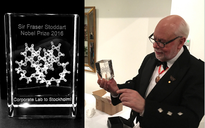
The photo below shows Sir Fraser Stoddart, winner of the Nobel Prize in Chemistry 2016, holding one of our blocks. Other Nobel prize winners we've produced blocks for include Sir John Walker, Sir Tim Hunt, Dr Edvard Moser and Dr May-Britt Moser, and many other eminent scientists.